
Бесплатный фрагмент - Is it possible to overcome aging?
Today and tomorrow cell therapy
R e v i e w e r s:
Vitaly Ivanovich Dontsov, Doctor of Medicine. Leading Researcher of the Federal information Center “Informatics and Management”, Laboratory “System Analysis and Information Technologies in Medicine and Ecology” of the Russian Academy of Sciences, Moscow;
Alexander Alexandrovich Zgurskiy, Doctor of Biology, CEO of “Medbiokor” LLC, Moscow.
Prokhorov, Leonid Yu.
Is it possible to overcome aging? Today and tomorrow cell therapy / Leonid Yu. Prokhorov. — Moscow/Ekaterinburg: “Publishing solutions” Ltd under license from “Ridero”, 2021. – 96 p.
2nd edition revised and expanded.
The book of Doctor of Biology L. Yu. Prokhorov describes the methods of modern cell biology that can be applied for rejuvenation of human skin using the person’s own cells. It briefly discusses the symptoms of aging, characteristics of the human skin and causes of skin aging, methods of obtaining and cultivation of autologous (self) skin fibroblasts, the procedure of cell therapy, results of the application of autologous fibroblasts. The book shows the need for long-term storage of cells in frozen form at low temperatures (-196ºС) in liquid nitrogen. It is anticipated that cell therapy can be used not only for skin rejuvenation but also to restore old or diseased organs, which will significantly increase the duration of human life.
This book also discusses some advances in cellular technology which, when implemented, will help to significantly improve rejuvenation of the skin of a person, as well as of other tissues and organs.
The book is intended for a wide range of readers, including students, postgraduates, researchers of biological and medical Universities, as well as for anyone interested in aging and rejuvenation.
Key words: longevity, rejuvenation, cell therapy, artificial organs, telomerase, old cells, young cells.
When designing the cover used the pictures from the internet.
© Prokhorov L. Yu., 2017
© Prokhorov L. Yu., 2021
© Design: “Publishing solutions”, Ltd, 2021
INTRODUCTION
One of the main urgent problems of humanity is old age and related problems: senile diseases, deterioration of appearance, atherosclerosis, dementia, carcinogenesis, ischemic disease, myocardial infarction, etc. What should we do? The answer is very simple: you must always stay young, periodically rejuvenate the aging body and then there will be no problems listed above. For most people, this is fantastic, but now there are methods that allow you to rejuvenate the skin. The next step is to rejuvenate other tissues and organs. It was found that the number of living, working and capable of dividing cells is constantly decreasing with age (Strehler, 1964). Dead cells are replaced by connective tissue. Connective tissue plays an important role in the aging process. In some aging organs, dead specialized cells are replaced with connective tissue cells that are unable to perform the functions of this organ. For example, cardiomyocytes (muscle cells of the heart) when they die as a result of a heart attack can be replaced by fibroblasts, forming a scar that is deprived of the ability to contract.
Therefore, it is important to replace old or dead cells with young, fully functioning ones.
Another task is to eliminate the old connective tissue and replace it with a young one. The use of both methods in combination can give the maximum effect.
A particular task that is already being solved today is skin rejuvenation. To rejuvenate the skin, eliminate wrinkles, scars after surgery, and acne, methods have already been developed that give an unambiguously positive result. This and some other topics related to the rejuvenation of human tissues and organs will be discussed in this publication.
EXTERNAL AND INTERNAL MANIFESTATIONS OF HUMAN AGING
With age, changes are occurring in the overall size, shape and composition of the body, soft parts of the face and skin, changes the bone system.
► Skin changes usually begin after 40 years, especially noticeable for the structure of the upper (epidermal) layer, which is thinned and flattened. By the age of 80 its thickness is 25% less than at the age of 30. The relief of the skin pattern on the palms, soles and fingers of hands and feet is smoothed. The changes affect the papillary layer of skin proper (dermis), which also becomes thinner. Gradually atrophy occurs in subcutaneous fat, which in long-livers may eventually disappear completely. Brown pigment spots appearing on the skin are characteristic for the back of the hands, shoulders, chest, etc. (Khrisanfova, 1999).
According to other data, the visible aging of the skin take place from the age of 25 years, when subcutaneous fat tissue begins to becom thin, and the elasticity of collagen structures decreases (Guide to gerontology, 2005).
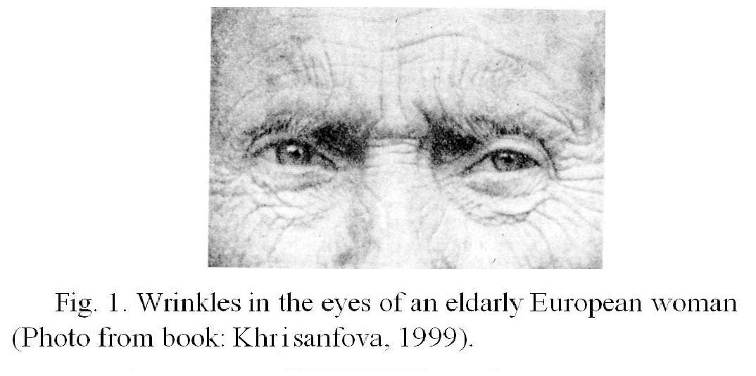
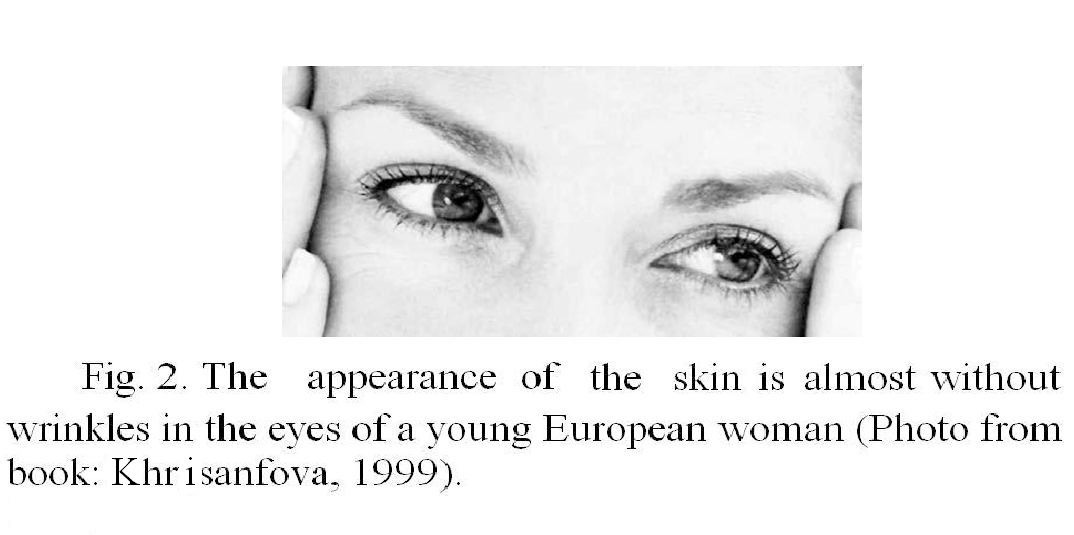
Due to the decrease in the number of sweat and sebaceous glands the skin becomes drier, its elasticity is lost. In men, the decrease in sebum occurs later than in women. The most obvious signs of aging are wrinkles. With age, there are multiple wrinkles on the skin, especially in open areas, such as the face, for example, the so-called «crow’s feet» at the outer corner of the eye. For fig.1 shows a picture of an elderly European woman, and in fig.2, for comparison, — photo of a young woman. The face ages faster than other parts of the body (Khrisanfova, 1999). This is explained by the fact that the person is more exposed to atmospheric influences, including sunlight, compared to other parts of the body. Aging under the influence of sunlight is called photo aging, since sunlight accelerates skin aging. Especially this process is affected by ultraviolet radiation, as part of the solar spectrum (Molochkov et al., 2005). The exposure to wind, dust, dryness, the action of microorganisms, detergent components, etc. accelerate skin aging.
The skin temperature decreases, especially among long-livers. This is due to a general decrease in metabolic processes, but partly due to the deterioration of blood supply and changes in sweat glands. Due to the decrease in their number, the excretory function of the skin is weakened (Khrisanfova, 1999).
► Significant changes are happening to the hair. Starting from the age of 30, the density of hair on the skin decreases (the density is a number of hair on a surface unit), they turn gray, that is, they cease to be colored with pigment. One of the reasons for graying is that the cells of the hair follicles (melanocytes) lose the ability to form a dye pigment. However, it is expected that another, more important reason is violation of a process of pigment transfer from melanocytes to the growing hair (Prokhorov, 2015b). Although hair growth is declining, older women often have facial hair (on the upper lip and chin). At the same time, hair on the body, limbs and eyebrows may disappear after 60 years.
► The bone system worsens, the curvature of the spine increases; intervertebral discs and cartilage of the articular surfaces of the bones flatten. A permanent sign of aging over the age of 45—50 years is bone thinning — osteoporosis.
► With age, there occur changes the overall size, shape and body composition, soft parts of the face.
► The height of a person decreases. According to some data, after 60 years, the height of men and women decreases by an average of 0.5−1.0 cm over a five-year period. This is due to the flattening of the intervertebral discs and increased stoop.
►There occur changes in the ratio of body components — muscle and fat. The amount of muscle tissue attains a peak at the age of 20—30 years, then its decline begins, at first slowly, and then with increasing rate, especially after 50 years. In this connection there occurs a decrease in muscle strength; at the age of about 70—80 years such strength reduced approximately two times. The subcutaneous fat layer decreases and the volume of internal fat in the abdominal area increases.
►Aging of the nervous system manifests itself in many ways. This applies to both the Central nervous system (brain) and the peripheral nervous system. Aging is manifested in functional and psychological changes that affect mental and physical performance, memory, emotions, complex behavioral reactions and other aspects of life. Structurally, aging is expressed primarily in reduced number of nerve cells — neurons. Although some reduction may occur already shortly after birth, a significant loss takes place in relatively late period, since the age 50—60 years, and such reduction is uneven in different areas of the brain. The degree of neuronal loss in the brain cortex of old people can reach as much as 40—50% or even more. In old age, neuronal density and size of neurons, diminish, together with deposition of pigment.
Age phenomen are also observed in the spinal cord and peripheral nervous system, they are observed in all parts of the vegetative nervous system (Khrisanfova, 1999).
— Aging worsens the condition and functions of other systems, tissues and organs of the body, including sense organs, cardiovascular, autonomic and immune systems, digestive organs, endocrine system, etc. (Guide to gerontology, 2005).
Now we can definitely affirm that senile changes may not be completely eliminated with diet, use of herbs, drugs, vitamins, physical exercise and the like.
How can we stop aging? The following sections will be described that the first step has already been done, and it is a cell therapy for a skin by its own cells. The next steps will be aimed at improving this technique and extending it to other tissues, systems and human organs.
CHARACTERISTICS OF THE HUMAN SKIN AND CAUSES OF SKIN AGING
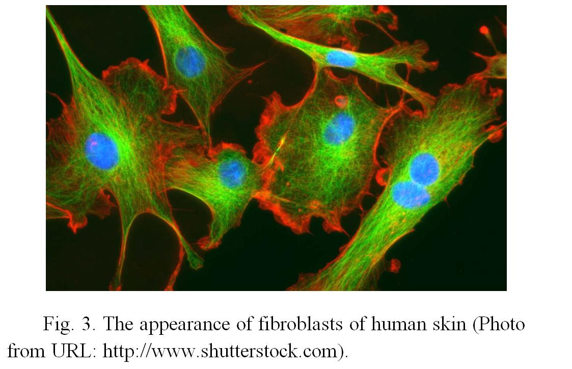
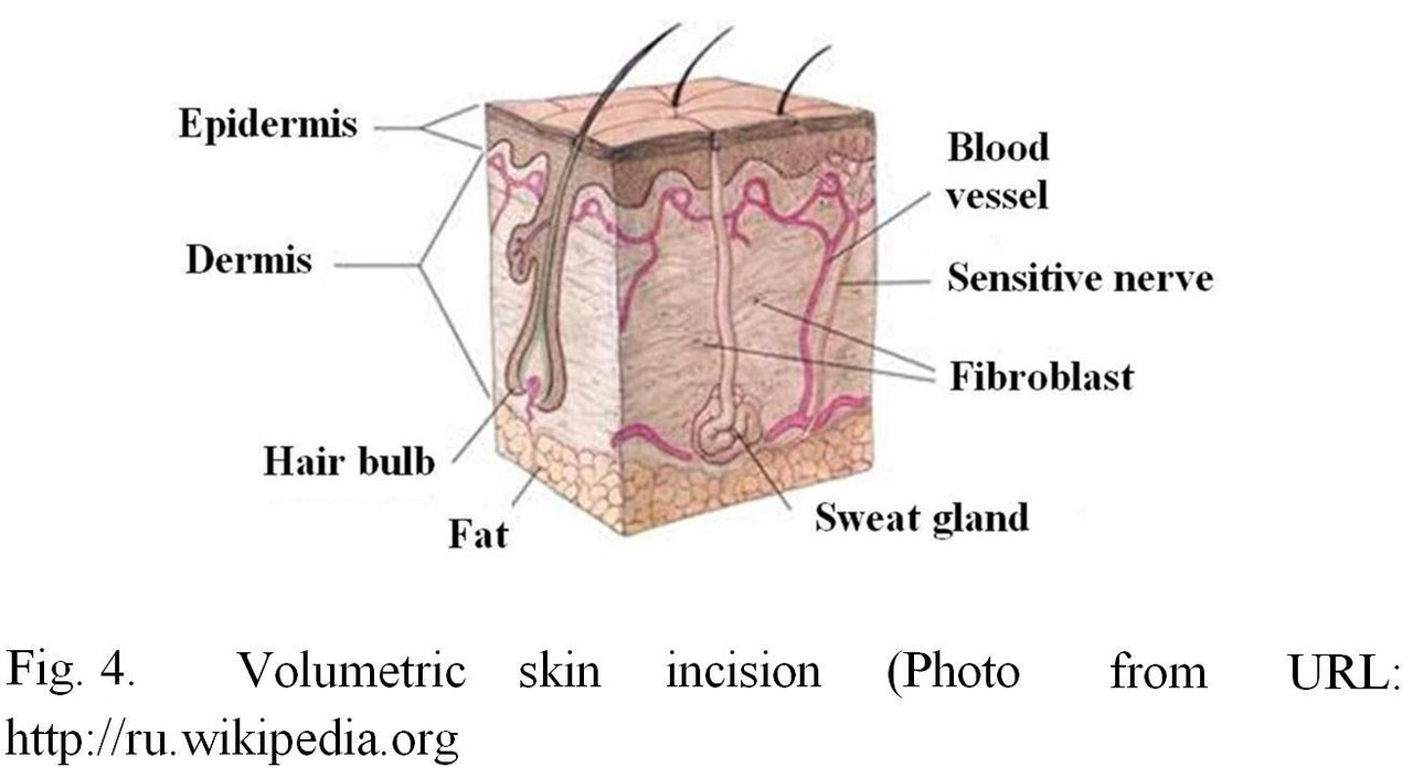
The main cellular elements of connective tissue are fibroblasts. Fibroblasts are cells of mesenchymal origin, round or elongated, spindle-shaped or with flat shape, spikes and oval nucleus (Fig. 3). The state and function of the main extracellular components of connective tissue: collagen, elastic and reticular fibers and intercellular matrix, depend on the functional activity of fibroblasts. As the result of differentiation of the fibroblasts become less active, mature cells — fibrocytes.
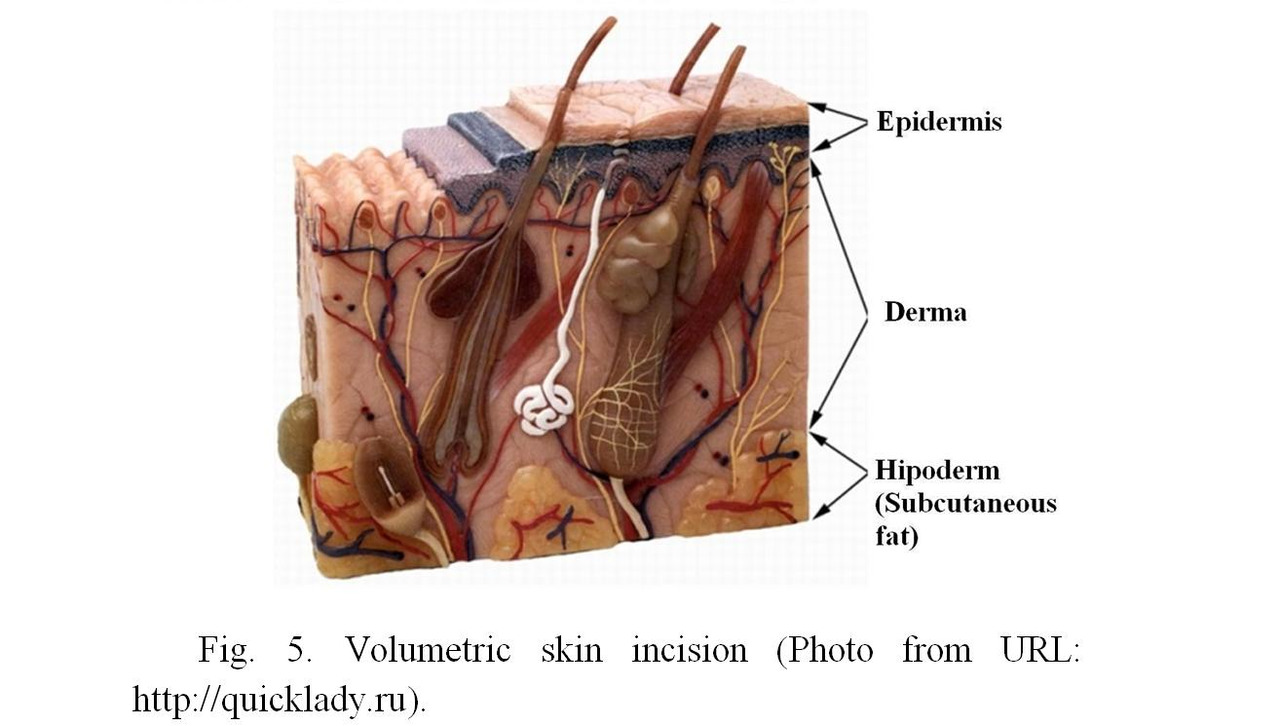
Fibroblasts are the main cells of the middle connective tissue layer of the skin, called derma (Fig. 4, 5). The principal function of fibroblasts in dermal skin layer is participation in metabolism of cellular substance. Fibroblasts of the skin synthesize and secrete into the environment a large number of biologically active substances, among which there are various growth factors, extracellular matrix components and enzymes. This process takes place continuously, and due to it intercellular substance is constantly replaced. Particularly intense is metabolism of hyaluronic acid (Kricheli et al., 2004). As in all other organs and tissues of the human body, there are age-related changes in the skin during the whole life.
In aging skin, the thickness of the dermis decreases, the moisture content in it falls, and as a result of this process the skin loses elasticity. The consequence is the formation of wrinkles. Aging of the skin in different parts of the body proceeds unevenly. Particularly rapid is the aging of exposed skin. The reason for this, as described above, is photo aging (because of exposure to sunlight), as well as atmospheric effects. At the same time, age-related skin changes are less pronounced in covered areas.
It is believed that one of the main causes of skin aging is a decrease in the ability of skin fibroblasts to divide with age. As a result of this process, the number of fibroblasts in the skin decreases, and they become less active. Therefore, if to stimulate proliferative and synthetic activity of fibroblasts, in any way, then it will be possible to improve skin conditions (Krikheli et al., 2004).
In addition, in the skin there occurs a decrease of the amount of collagen fibers and their hydration, they lose the ability to swell and become rigid; as a result the turgor is decreased, and wrinkles appear. At the same time, the skin loses lipids, its protective mantle breaks down, and it becomes vulnerable to disruption (Guide of gerontology, 2005).
As the number and activity of fibroblasts is reduced, the remaining cells are unable to compensate for senile changes in the skin. Therefore, if to increase the number of young active fibroblast cells in the skin, it is reasonable to wait for the improvement of its condition due to the fact that fibroblasts will produce young collagen and elastin, and this will increase the elasticity of the skin and its turgor; as a result, the wrinkles well be smoothed.
METHODS OF SKIN REJUVENATION
In order to correct age-related changes in the skin, there is a variety of currently used methods (peels, polishing, lifting, etc.) and preparations (toxin of botulism, components of extracellular matrix and connective tissue, etc.). Many of these methods are aimed at stimulating skin cells, in particular fibroblasts. However, it can be possible to solve the problem in
another way — by increasing the number of fibroblasts in the skin by means of transplanting them to the places where needed.
Before transplantation of fibroblasts became a reality, researchers had to solve many methodological questions, including those of safety of similar procedures.
Significant progress has been achieved in this regard when researchers became able to cultivate the cells, i. e. maintain the viability of fibroblasts outside the body, or in other words in vitro. In 1961, L. Hayflick and P. S. Moorhead (Hayflick, Moorhead, 1961) reported that even under optimal conditions of in vitro cultivation, human embryo fibroblasts were only capable of dividing a limited number of times (50 ± 10). In subsequent studies, this observation was repeated many times. The last phase in the life of cells in culture hase been identified as cellular senescence, and the phenomenon received the name of the author as Hayflick limit.
After the establishment of Hayflick limit and as a result of numerous studies, it was found that normal fibroblasts in culture retain a diploid karyotype and have a limited life expectancy. In addition, normal cells lack oncogenic potency. These requirements to cultures of normal cells were issued in the form of normative documents.
If these requirements are met, it is possible to use human fibroblasts cultivated outside the body for the production of immunobiological prepartions, and then for therapeutic purposes. Scientific research and clinical developments in this area are very intensive, due to general rise of cellular technologies based on stem cells.
Currently, there are two main approaches to the treatment of skin with preparations containing live fibroblasts. On the one hand, methods and preparations for the treatment of skin defects due to wounds and burns became widely known with the help of cultures of allogenic (from another organism) embryonic fibroblasts. An alternative to these methods is the possibility of using autologous (a person’s own) human fibroblasts for cell replacement therapy of the skin (Zgursky, 2004).
It is believed that the best results of cell therapy are obtained by using cultures of embryonic fibroblasts, which have a greater proliferative potential than cultures from adult donors. However, the use of cells derived from embryos has a number of limitations, including ethical ones. At the same time, data accumulate that a person’s own fibroblasts retain their potential during aging, despite a decrease in their number in the aging organism (Terekhov, 1984).
To achieve primary results, it is necessary to obtain fibroblast cultures from adult donors. It was found that proliferative abilities of the fibroblasts obtained (in comparison with embryonic skin fibroblasts) are sufficient to use them for cell therapy (Zgursky, 2004).
Methods of obtaining and cultivation of autologous skin fibroblasts
Cell material to obtain a culture of autogenous fibroblasts is taken by biopsy of the skin (a small section of the skin with a size of about 0.5 cm2) in the forearm or in the ear area of the patient with local anesthesia.
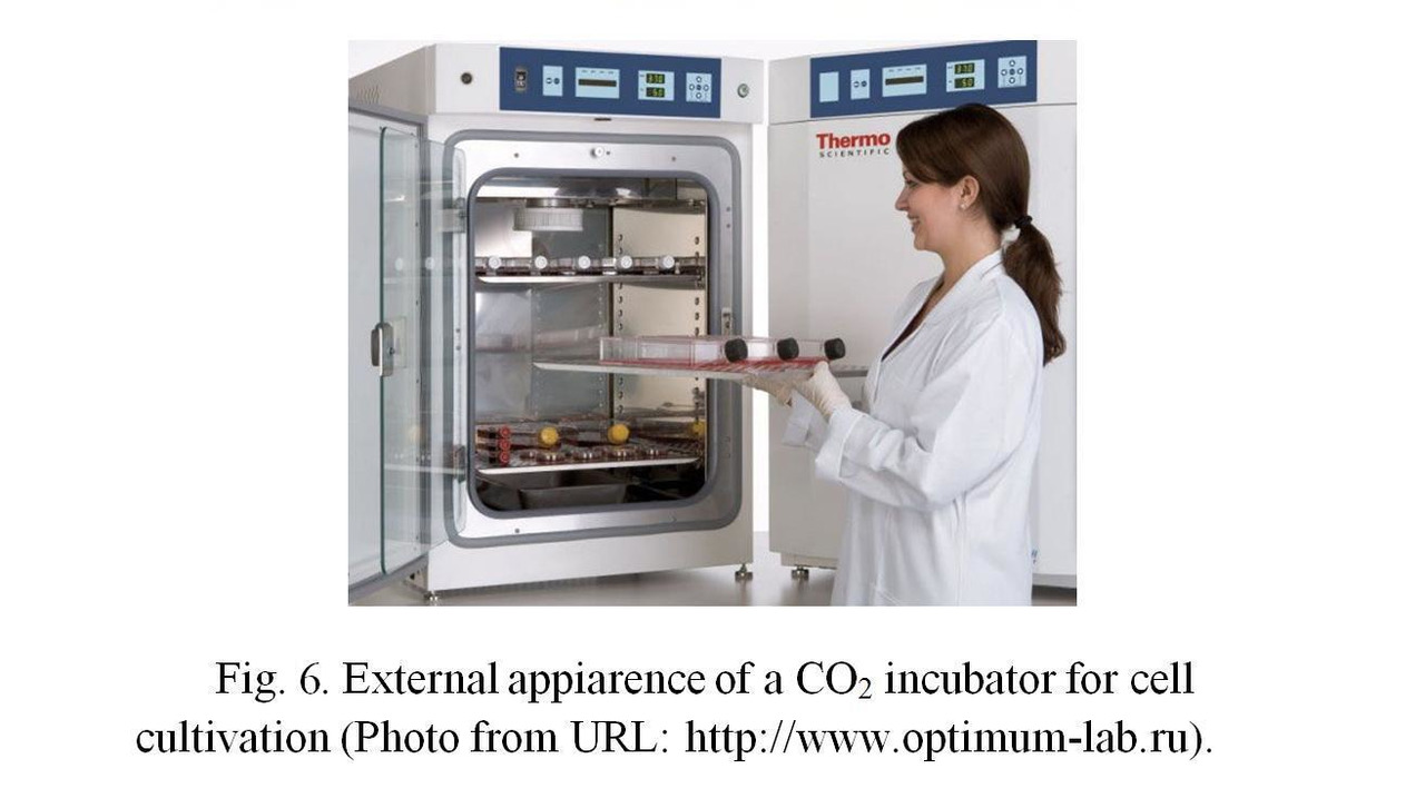
Obtaining and cultivation of fibroblasts from skin biopsy are carried out according to the standard procedure. To obtain the primary culture of cells, the tissue is washed with a physiological solution containing antibiotics, placed in a sterile Petri dish and treated with a solution containing trypsin or collagenase (both are enzymes). Under the influence of this solution, the tissue softens, and living cells (fibroblasts) spread from it to the bottom of a dish. Then the dish is filled with nutritive growth medium (see. below) and placed in a CO2 incubator (special tightly closed box that maintains a constant temperature of 370C and an increased concentration (5—7%) of carbon dioxide in the air (Fig. 6); the usual concentration of CO2 in the atmosphere is 0.03−0.04%, in which fibroblasts cannot grow). Cells begin to multiply and after filling the whole bottom of a dish, the fibroblasts are removed from the growth surface with a mixture of solutions: Versene (solution of EDTA-ethylene diamine tetraacetic acid and inorganic salts) and trypsin. The primary cultured cells are centrifuged, washed from the solution components and suspended in the culture medium.
Next, the cells are transferred into culture flasks (Figures 7a, 7b), containing liquid growth medium.
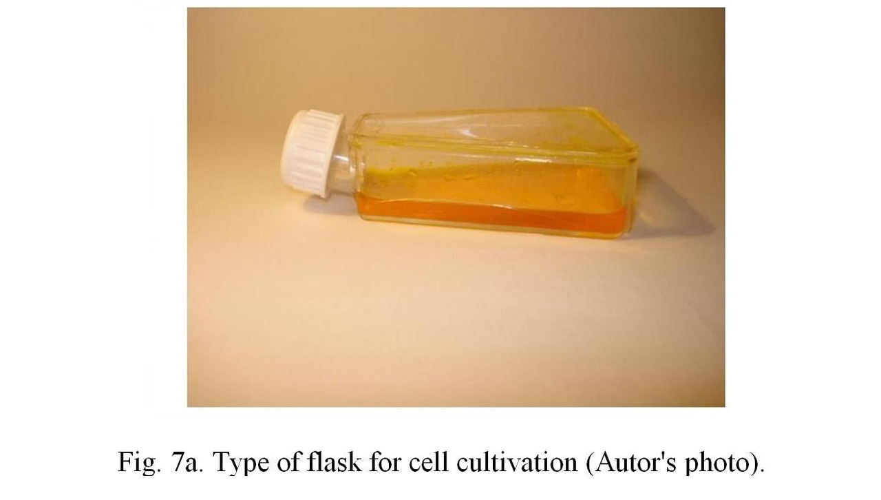
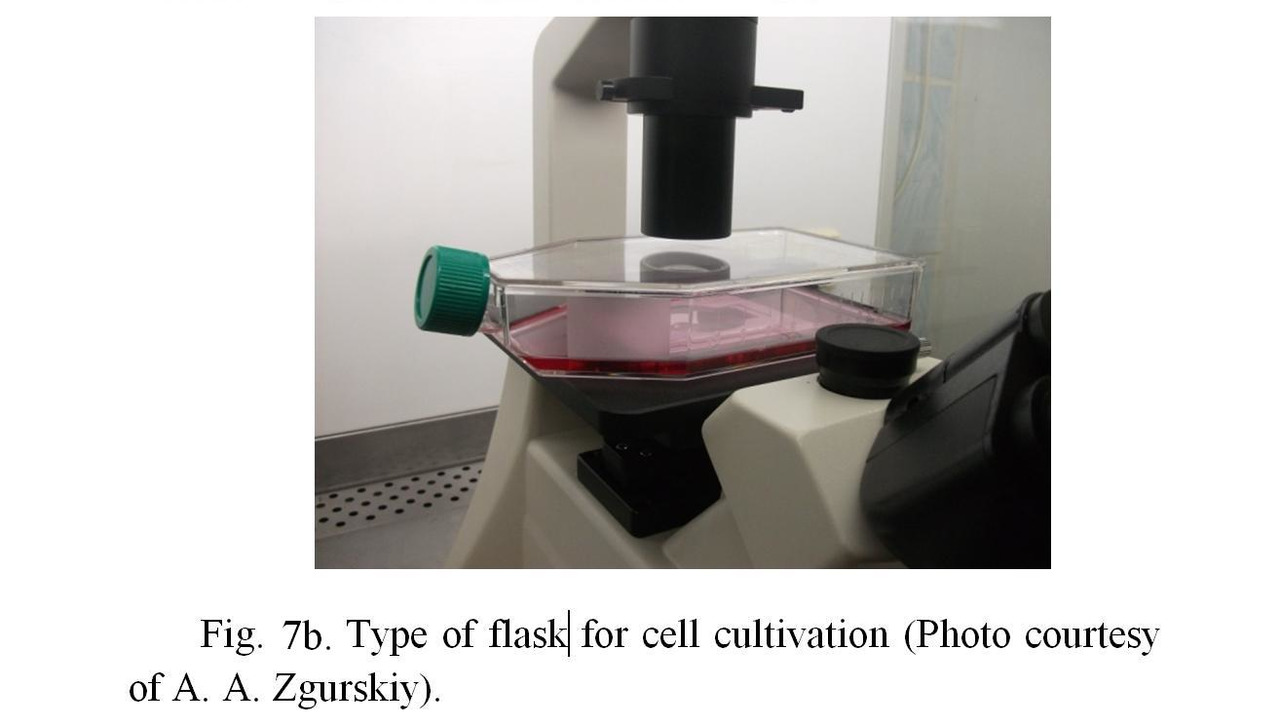
Growth medium contains a set of amino acids, glucose and others nutrients, as well as 10—20% fetal calf serum containing the necessary growth factors for cell division. The flasks themselves are placed in the CO2 incubator. Flasks do not closed tightly, so that the air with a high content of carbon dioxide from the incubator could flow into the flasks. It is possible also to use flasks with ventilated lids (with built-in sterile filter), through which an exchange with air in the incubator is performed. This method provides conditions for the growth (division) of cells.
All works are carried out in strictly sterile conditions in a laminar flow hood (Figures 8a, 8b), in which the fan pumps sterile (free from bacteria) air through the sterilizing air filter, located at the back or top of the box, with a small additional pressure.
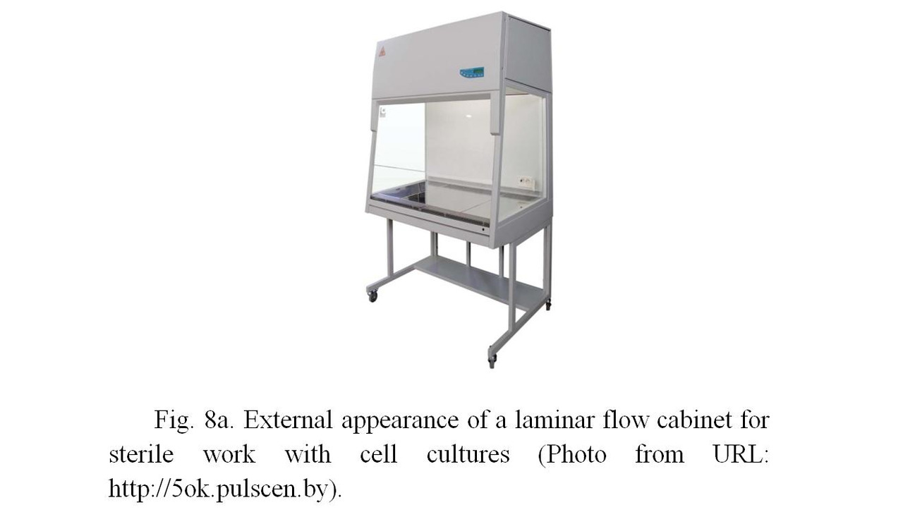
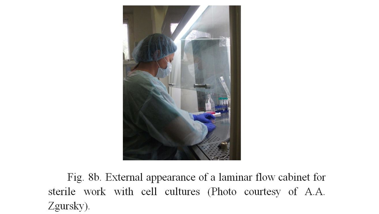
Due to excessive pressure inside the laminar hood, the external air with bacteria does not penetrate inside the box, so cell cultures remain sterile. The work inside the laminar hood is carried out by a highly qualified specialist using sterile laboratory glassware, including flasks, pipettes, Petri dishes, etc. Additional sterility is carried out by firing the surfaces of flasks neck, pipette working parts in the flame of an alcohol or gas burner, which is installed inside the laminar hood and is turned on constantly during the entire period of work with biological material. The specialist himself is outside the laminar hood, but all actions are carried out by hands in sterile surgical gloves inside the hood through an open window, from which the sterile air constantly leaves, what does not allow the non-sterile air (with bacteria) from the outside.
Cell cultures obtained from patients are used to develop the necessary number of cells sufficient for procedures (6—8 passages or cell divisions in the culture). In further procedures the fibroblasts are tested for absence of contamination (infection) of HIV/AIDS, hepatitis A, B and C, mycoplasma and chlamydia. Testing is carried out using PCR (polymerase chain reaction) and immunoassay methods. The lack of oncogenic potential of cells is determined on athymik (non-immune) mice. Next, a monolayer culture of cells is removed from the surface of the flasks with a mixture of solutions of Versene and trypsin and suspended (stirred) using serological pipettes in sterile saline for injection. Then the cells centrifuged and washed 2—3 times from Versene and trypsin in the same saline. The final suspension is stored at +40C. Cell preparations are used within 24 hours after obtaining the suspension.
The viability of cells in the suspension is determined by establishing the culture after different periods of incubation and counting the number of attached cells, as well as their ability to form colonies.
The procedure of cell therapy
The procedure for administration of cells to the patient is carried out by means of tunneling injections for large wrinkles or multiple injections for small wrinkles.
The number of cells in the preparations varies depending on the procedure, the volume of the preparation administered is on average of 1—3 ml per procedure. The number of injected cells is on average from 1 to 10 million per procedure.
The method of administration of cells is shown in figures 9a, 9b. Cells are injected using an insulin syringe into the dermal (middle) layer of the skin (Zgurski, 2004). The needle of insulin syringe is very thin, so the procedure causes minimal discomfort.
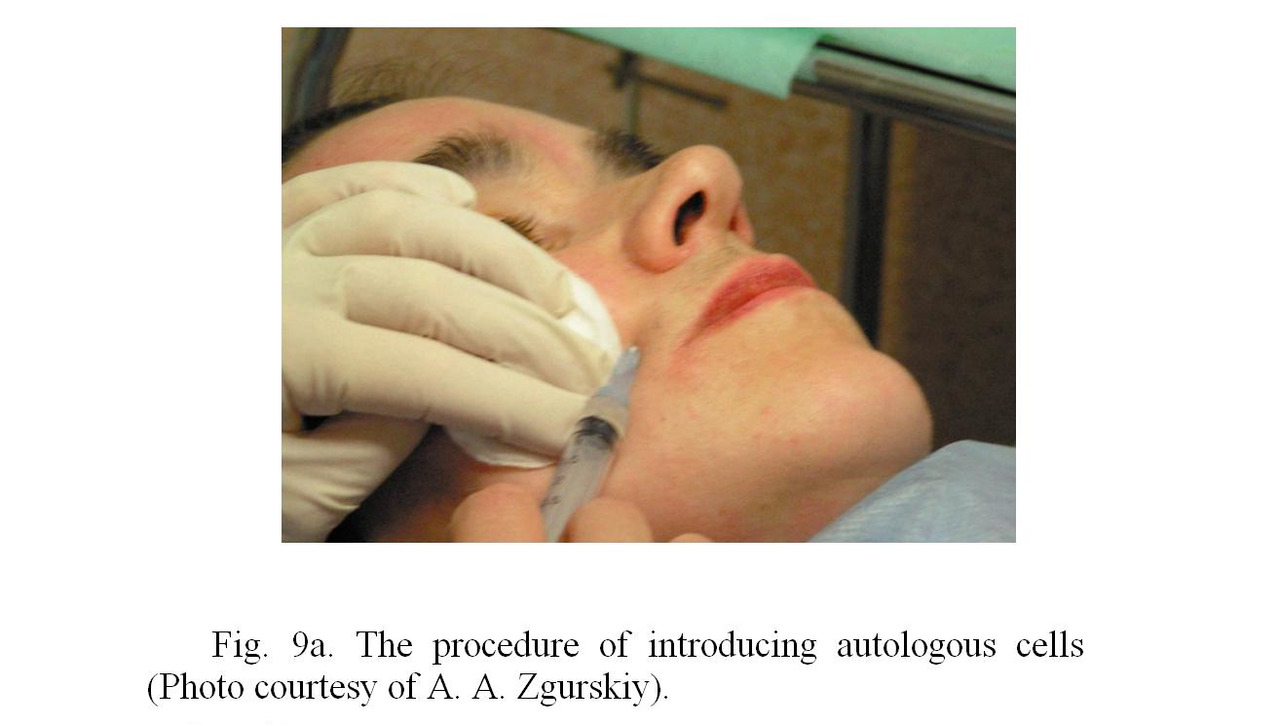
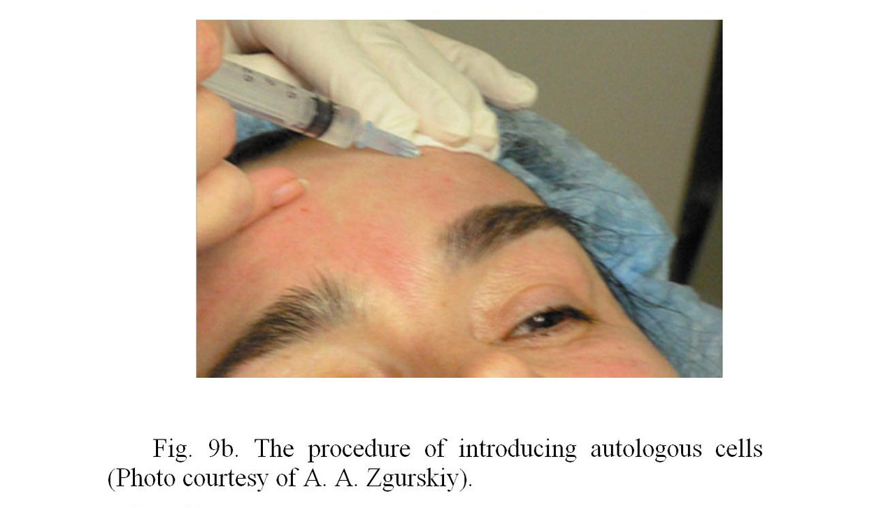
The surface of the skin into which the cells are injected is treated with sterilizing solutions before performing the procedures, and all the manipulations are performer strictly in sterile rubber gloves and in a gauze bandage covering the respiratory organs.
The results of the application of autogenous fibroblasts
According to one of the leading experts in the application of autologous fibroblasts, the PhD of biological Sciences A. A. Zgursky, the procedure has several attractive aspects for cosmetologist and, accordingly, for the patient (Zgursky, 2004):
— it uses the patient’s own cells;
— a single biopsy is usually performed;
— there is only a slight discomfort in the process of applying procedures for the patient;
— the effect lasts for a long time;
— easiness of application;
— a small number of procedures are performed to obtain the effect;
— there are no complications and side effects.
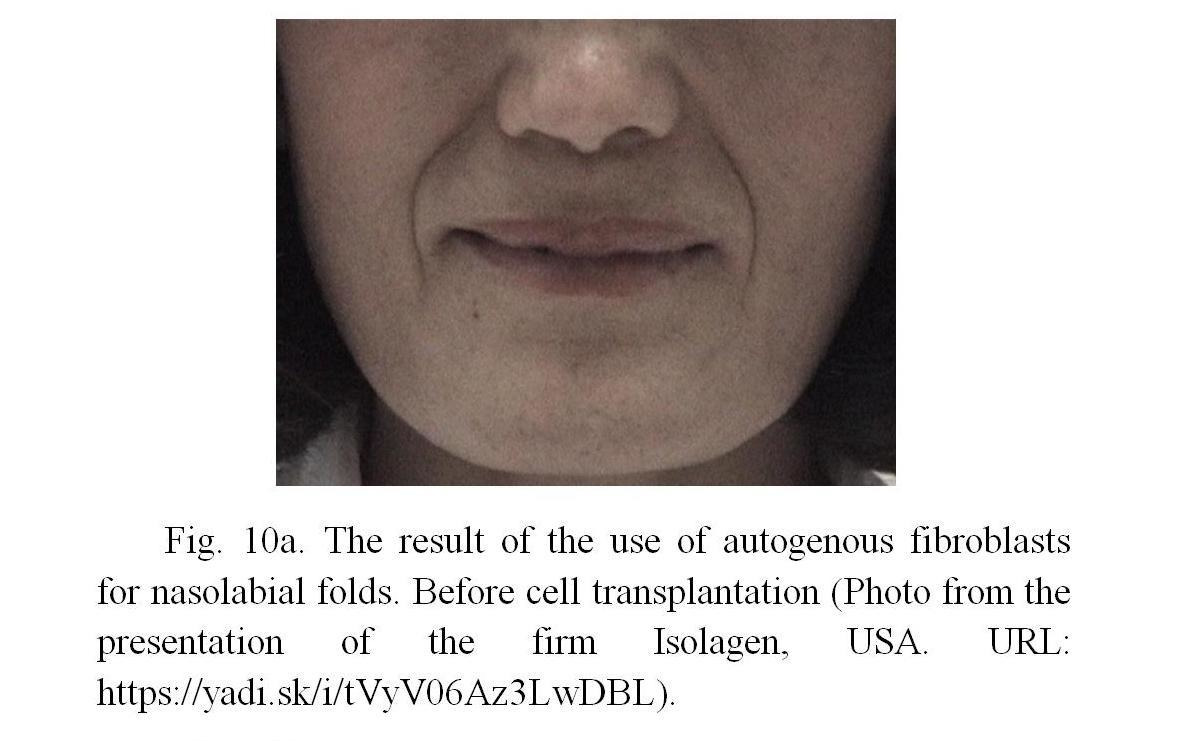
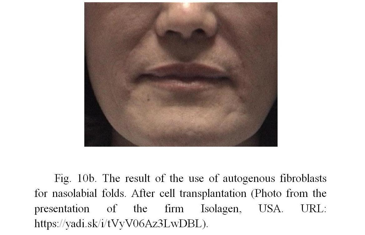
Autologous fibroblasts are used for the correction of wrinkles, atrophic scars, atrophic areas of the skin, disorders of skin structure (Boss et. al., 2000).
Бесплатный фрагмент закончился.
Купите книгу, чтобы продолжить чтение.